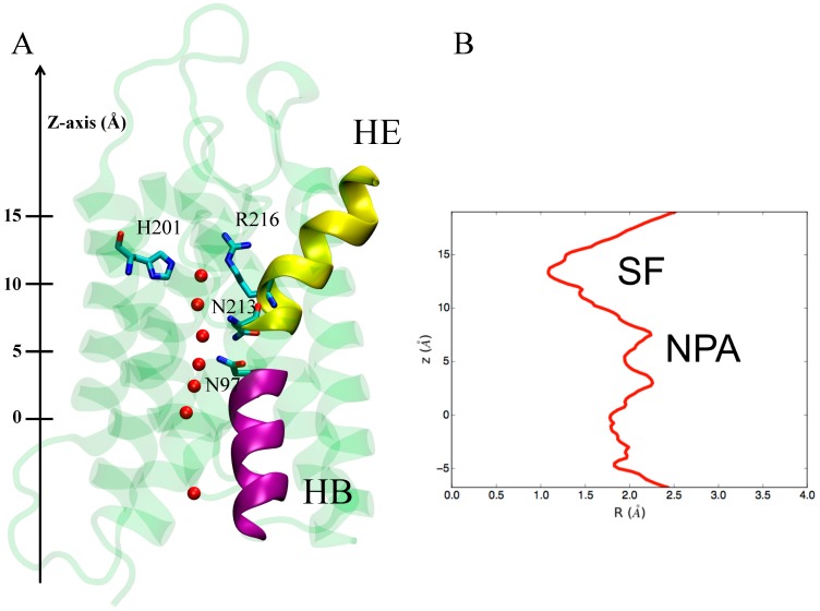
Figure 2.
(A) X-ray structure of hAQP4. The X-ray solved structure of hAQP4 (PDB code 3GD8 [21]) is depicted as green cartoon representation. Important residues in the constricted selectivity filter (H201 and R216) and the asparagine residues belonging to the NPA motif regions are rendered as sticks. Water molecules inside the pore are shown as red spheres. The two short pore alpha helices responsible for an electrostatic barrier preventing proton conduction and named HE and HB are depicted as yellow and magenta cartoon representation respectively; (B) Pore radius. Pore radius R(Å) profile along the z-axis obtained from the hAQP4 X-ray structure using HOLE as cavity detection software (Department of Crystallography, Birkbeck Collage, University of London, London, UK) [51].

