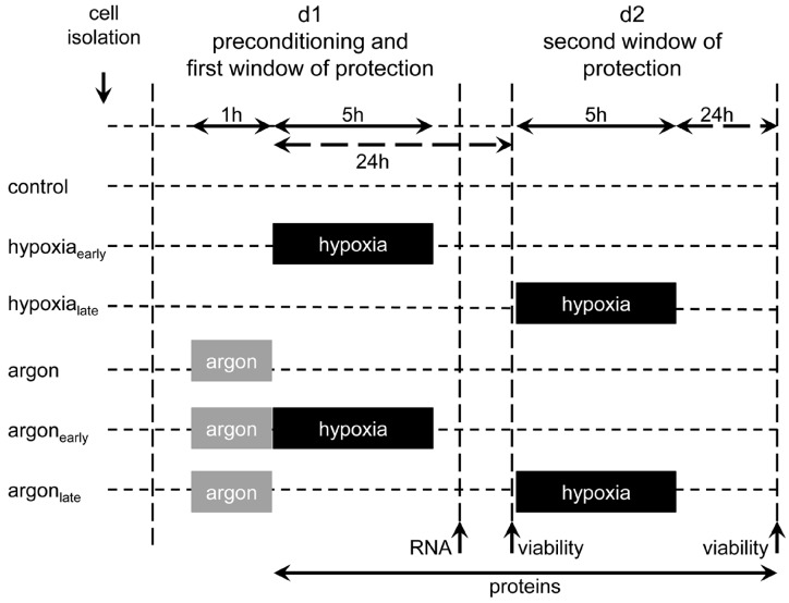
Figure 6.
Experimental set-up. Isolated rat heart cells, pure cardiomyocytes, and fibroblasts were subjected either to 50% argon (50% Ar, 24% N2, 21% O2, 5% CO2) or room air (74% N2, 21% O2, 5% CO2) for 1 h at 37 °C. Cells were either challenged within the first (0–3 h) or second window (24–48 h) of preconditioning with an ischemic insult of 5 h. Cell survival was assessed 24 h after prolonged hypoxia via MTT assay. To unravel the underlying mechanisms, mRNA expression was analyzed 8 h after argon treatment. For investigating the activation of pro-survival kinases, cell lysates were prepared at different time points (0 h, 0.5 h, 4 h, 8 h, 24 h, 48 h) after preconditioning.

