
Figure 5. Epithelial adhesion, anti-phagocytosis and virulence of D39s and its cps promoter replacement derivatives.
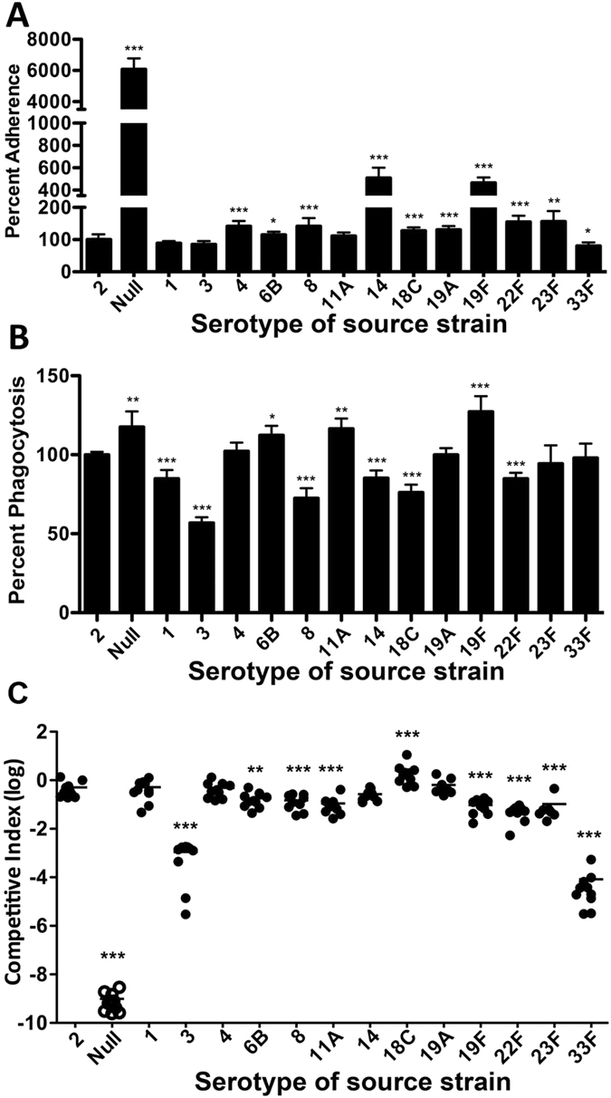
(A) Pneumococcal adhesion to human alveolar epithelial A549 cells. D39s and its promoter replacement derivatives were incubated with the confluent monolayers of A549 cells for 1 h before the monolayers were lysed to enumerate the number of viable pneumococci on TSA blood plates. The adhesion levels of the promoter replacement strain are displayed as the percentage values relative to the value in D39s. (B) Phagocytosis of the cps promoter derivatives by RAW264.7 murine macrophages. The RAW264.7 cell monolayers were infected with the pneumococci, treated with antibiotics, and lysed to quantify intracellular (phagocytosed) bacteria as in A. The phagocytosis level of each promoter replacement strain was shown as the percentage value relative to the value of the parent strain D39s. (C) The virulence levels of the promoter replacement strains. CD1 mice were intraperitoneally infected with a mixture of D39 and one of the promoter replacement strains in a 1:1 ratio. The pneumococci in the bloodstream of the mice were enumerated 21 h post infection as in A. Each filled circle represents a relative bacteremia level or competitive index (CI) value of a single mouse. The open circles indicate the mice, from which no unencapsulated pneumococci were recovered. The horizontal bar indicated the mean in each group of mice. The parental strain (type-2) was used as a reference. *p < 0.05, **<0.01 and ***< 0.001.