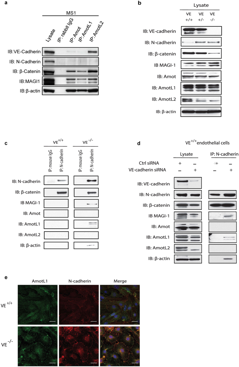
Figure 4. AmotL1 is recruited to N-cadherin associated adhesion complexes in the absence of VE-cadherin.
(a) Co-immunoprecipitation was performed using antibodies against Amot, AmotL1 and AmotL2 in MS-1 cells. AmotL2 was shown to be associated with VE-cadherin whereas AmotL1 could not be immunoprecipitated with VE-cadherin or N-cadherin. (b) Western blot analysis of junctional protein expression in VE-cadherin+/+, +/− and −/− endothelial cells (c) Co-immunoprecipitation of N-cadherin with AmotL1 in VE-cadherin−/− endothelial cells. (d) Immunoprecipitation analysis of endothelial cells siRNA depleted of VE-cadherin (e) Immunofluorescent staining of AmotL1 (in green) and N-cadherin (in red) in VE+/+ and VE−/− cells showing co-localization in cellular adhesion junctions in VE-cadherin deficient endothelial cells. Nuclei were visualized by DAPI staining (in blue). Size bar, 10 μm.

