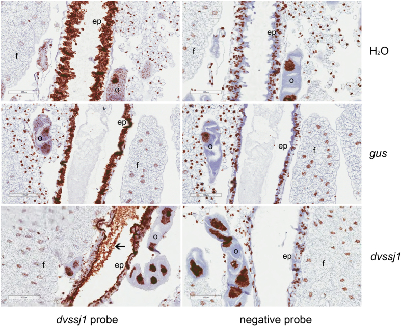
Figure 4. Visualization of dvssj1 mRNA expression in WCR 3rd instars by in situ hybridization.
Representative midgut sections (Supplementary Fig. 3) were from WCR 3rd instars treated with H2O (top panel), gus dsRNA (middle panel) and dvssj1 frag1 (bottom panel) at 50 ng μl−1 for 48-h. All treatments were hybridized with the dvssj1 probe and an RNAscope® negative control probe (Bacillus subtilis dihydrodipicolinate reductase (dapB) gene). Expression of dvssj1 mRNA is observed in midgut epithelium cells (ep) and oenocyte cells (o) of H2O and control gus treatment. Knockdown of dvssj1 mRNA in midgut epithelium cells (ep) and oenocyte cells (o) is observed in larvae treated with dvssj1 dsRNA (bottom panel). No clear presence of dvssj1 mRNA in fat body cells (f) and dvssj1 dsRNA in midgut lumen was observed (arrow). Images were captured at 40× magnification with 100 μm scale bars.

