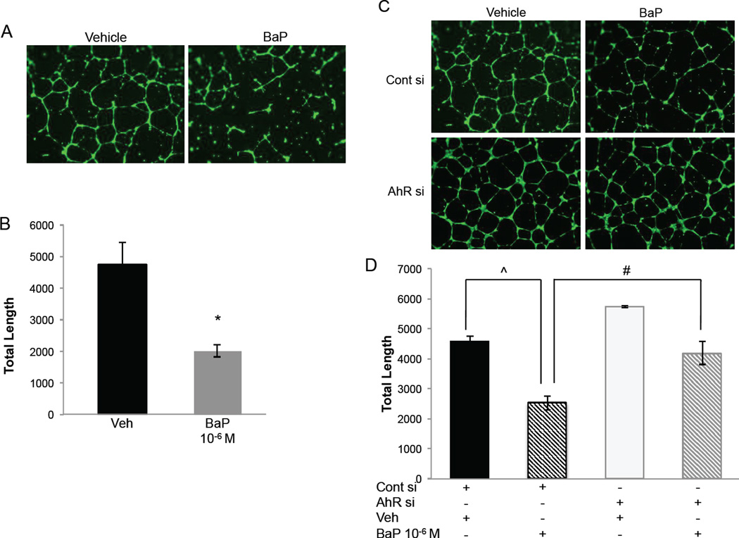
Figure 4.
BaP exposure results in impaired EC tube formation. (A) Representative tube formation images of vehicle- and BaP-treated endothelial cells. (B) Graphical representation demonstrates that total tube length is diminished in the setting of BaP treatment (*p=0.003). (C) Representative tube formation images in the setting of control siRNA and AhR siRNA transfections demonstrate that BaP-mediated effects on tube formation is mediated via AhR. (D) Graphical representation of total tube length shows that BaP treatment results in impaired angiogenesis in comparison to vehicle treatment in the setting of control siRNA transfection (^p=0.01). In contrast, when AHR is knocked down, tube formation is rescued in the presence of BaP (#p=0.008).

