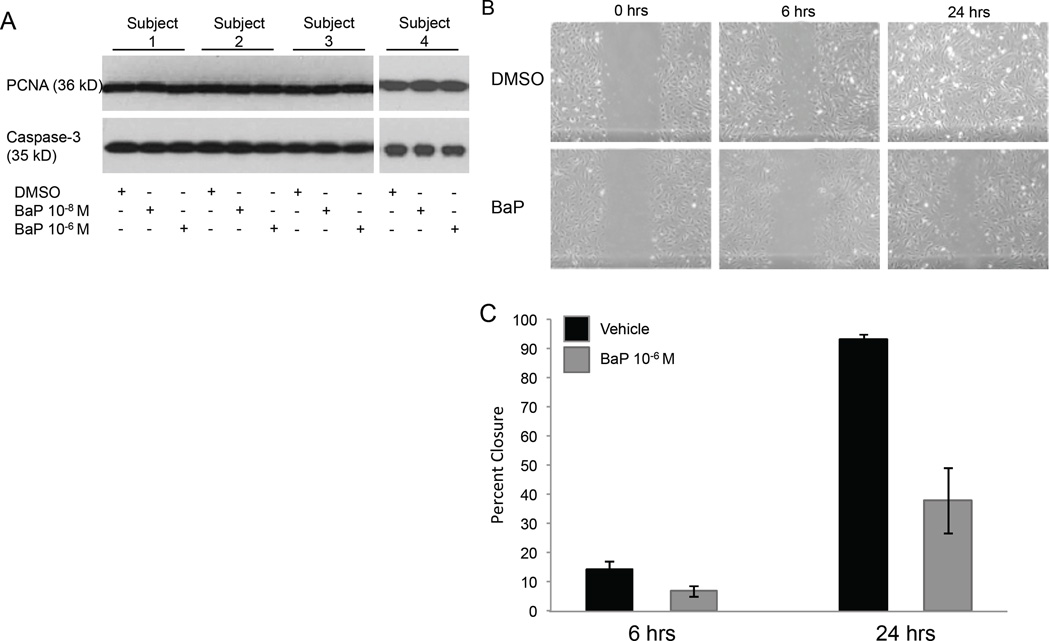
Figure 5.
Deficient EC tube formation is the result of impaired EC migration. (A) Representative western blots shows no difference in proliferation or apoptosis with varying concentrations of BaP treatment in all four subjects. (B) Representative wound scratch images from one subject demonstrate impaired migration after BaP treatment. (C) Graphical representation of wound scratch assays performed on all subjects (χ2 = 15.9, p=0.007).

