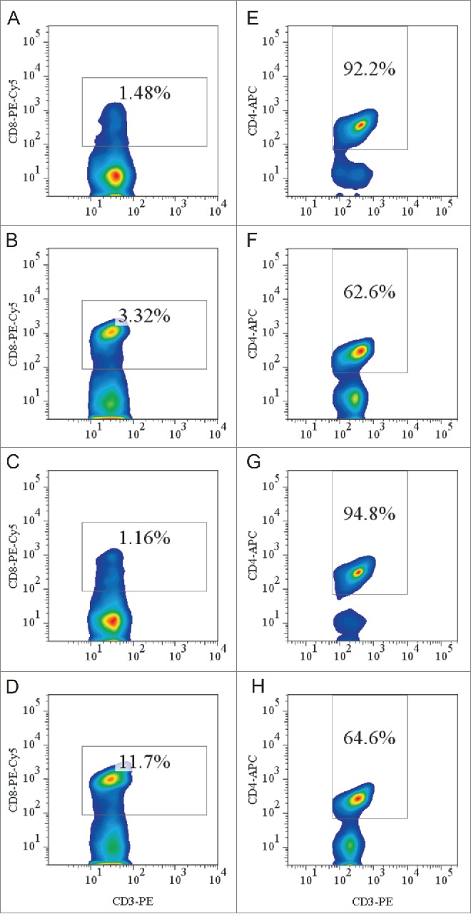
Figure 3.

Flow cytometric analysis of peripheral CD8+ and CD4+ (T)lymphocytes. (A-D) Splenocytes from HLA-A11/DR1 mice (A), HLA-A11 mice (B), HLA-DR1 mice (C), and wild-type C57BL/6 mice (D) were isolated. CD3+ T lymphocytes were gated by staining with an FITC-anti-CD3 mAb and CD8+ T lymphocytes were gated by staining with a PEcy5-anti-CD8 mAb. (E-H) Splenocytes from HLA-A11/DR1 mice (E), HLA-A11 mice (F), HLA-DR1 mice (G), and wild-type C57BL/6 mice (H) were isolated. CD3+ T lymphocytes were gated by staining with a PE-anti-CD3 mAb, and CD4+ T lymphocytes were gated by staining with an APC-anti-CD4 mAb.
