
Figure 3.
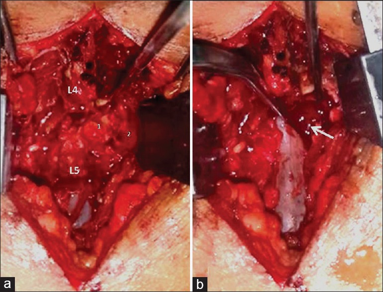
(a) Intraoperative photographs showing the ligamentum flavum (LF) (1) extending anteriorly beyond the facet joint (2) to get attached to the posterolateral aspect of L4 vertebral body. (b) Large epidural venous varices on retracting the dura after excision of LF (arrow)