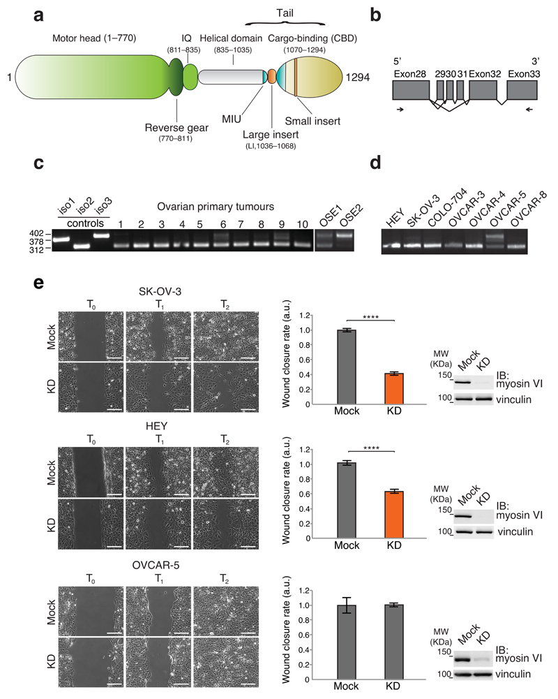
Figure 1. Myosin VIshort is selectively expressed in ovarian cancer cells and is critical for cell migration.
(a) Myosin VI domain structure. (b) Schematic representation of the region amplified by RT-PCR. Coding exons are represented by grey boxes. Alternative splicing events are depicted. Oligos used for the PCR mapped in Exon 28 and Exon 33 and are indicated by arrows. (c,d) RT-PCR from cDNA prepared from the indicated primary cells (c) and cell lines (d). Controls are from plasmids carrying the tails of the different isoforms. (e) Wound healing assay. The indicated cell lines were knocked down for myosin VI (KD) or mock treated. Left panel, sample images: T0 first frame, T1 and T2 arbitrary points identical for control and KD of the same cell line. Scale bars, 200μm. Central panel: quantification of the wound closure speed relative to control. Error bars, s.d. (n=6 movies for SK-OV-3 and HEY, n=8 movies for OVCAR-5. Data are from three independent experiments). **** P<0.0001; ns, no significant difference by two-tailed T-test. Right panel: anti-myosin VI immunoblotting (IB) performed at T0.

