Figure 3.

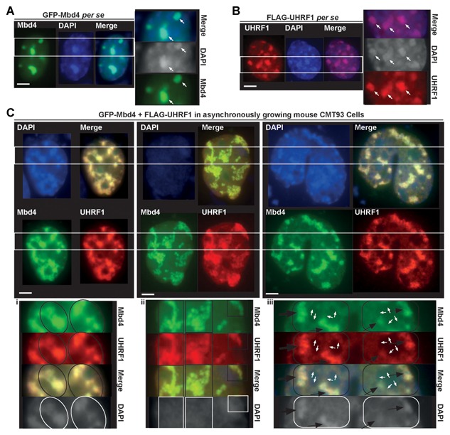
MBD4 tightly co‐localizes with UHRF1 at chromocenters. Asynchronously growing mouse CMT93 cells were grown on coverslips and transfected by GFP‐MBD4 (A), FLAG‐UHRF1 (B), or co‐transfected with GFP‐MBD4 and FLAG‐UHRF1 (C). The cells were fixed 48 h later and analyzed directly by immunofluorescence (IF) (A), or immunostained with anti‐FLAG rabbit primary and goat anti‐rabbit IgG‐Alexafluor Red conjugate secondary antibodies and analyzed (B & C). Nuclear counterstaining was visualized with DAPI. Scale bars, 10 µm. (A) Distribution of GFP‐MBD4 in CMT93 cells. (B) Distribution of FLAG‐UHRF1 in CMT93 cells. (C) GFP‐MBD4 and FLAG‐UHRF1 exclusively colocalized with each other at chromocenters in CMT93 cells. In the immunostaining images in (C. i & ii), the cells exhibit an increase in cell size and marked large‐scale reorganization of heterochromatin, which may be indicative of heterochromatin reformation and replication in interphase. In (C. iii), the cells appear to be undergoing orderly division into two daughter cells. The insets on the right (A & B) or below (C. i, ii, iii) correspond to magnifications of the areas indicated by the two parallel white line. Scale bars, 10 µm.
