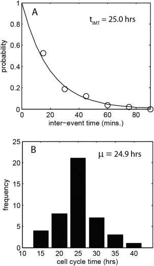
Figure 4.

A: probability distribution of the interevent time for events shown in Figure 3A, occurring between the 0 and 18 h time points (circles – measurement data, solid line – statistical fit). The t IMT value indicated is obtained by a best fit to the data assuming a nonhomogeneous poisson process. B: Histogram of measured cell cycle time obtained from manual, frame‐by‐frame tracking of 44 cells through their cycle.
