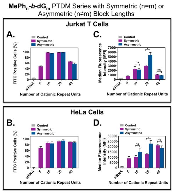
Figure 2.
FITC-siRNA internalization into Jurkat T cells and HeLa cells using symmetric and asymmetric ROMP-based protein mimics. Jurkat T cells (cell density = 4×10⁵ cells/mL) treated with polymer/FITC-siRNA complexes with an N:P ratio = 8:1 in complete media for four hours at 37°C and compared cells only receiving FITC-siRNA. HeLa cells (cell density = 5×10⁴ cells/mL 48 hours prior to experiment; 70–90% confluent on the day of the experiment) treated with polymer/FITC-siRNA complexes with an N/P ratio = 4/1 in complete media for four hours at 37°C and compared cells only receiving FITC-siRNA. All data was compared to an untreated control. A) Percent positive Jurkat T cells. B) Percent positive HeLa cells. C) MFI of the Jurkat T cell population. D) MFI of the HeLa cell population. Each data point represents the mean ± SEM of three independent experiments. * = p < 0.05 and ns = not significant, as calculated by the unpaired two-tailed student t-test. Statistics represents significance between symmetric and asymmetric block copolymer PTDMs with the same cationic charge block length.

