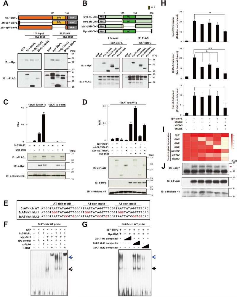
Figure 5. Dlx factors mediate Sp7's regulatory action in osteoblast gene regulation.
(A, B) Western blotting of co-immunoprecipitates of ectopically expressed indicated Myc-Dlx5 and Sp7-BioFL forms (upper panel) from 293T cell nuclear extracts.
(C, D) 3T3 cell luciferase reporter assay for indicated reporter constructs following transfection with indicated Sp7 and Dlx5 expressions. Data show the means ± SDs from triplicate experiments.
(E, F, G) EMSA of Dlx5 and Sp7 complexes with the AT-rich motif. Black arrow: Dlx5-DNA complex; blue arrow: Sp7/Dlx5-DNA co-complex.
(H, I, J) Sp7-FLAG ChIP-qPCR (H), RT-qPCR (I) and western blotting (J) analysis in MC3T3E1 cells following lentiviral transduction of shRNA-mediated knock-down of Dlx3, 5 and 6 and retroviral transduction of Sp7BioFL expression. The color scale indicates relative gene expression values in the qPCR analysis. Red arrow: Sp7-BioFL protein; black arrow: endogenous Sp7 protein. Data displayed are the means ± SDs from triplicate experiments. *: p < 0.05, **: p < 0.01.

