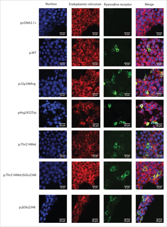
Figure 3.
Co-localization of RyR1 with the endoplasmic reticulum. Immunofluorescence of transiently transfected HEK293T cells with RyR1 cDNA. Variants are indicated by their amino acid change. Primary antibodies that specifically recognize RyR1 (34C) and PDI as well as fluorescently-labeled secondary antibodies FITC (green) and TRITC (red) were used to visualize RyR1 and PDI respectively, while nuclei were visualised by staining the cells with DAPI (blue). Cells were examined by confocal fluorescence microscopy at a magnification of 1260 X; the scale bar in each merged image represents a length of 20 microns.

