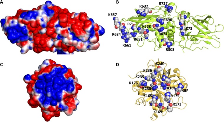
Fig 2. The putative anion-binding allosteric site of human FXIIIa.
(A) The electrostatic potential of the surface exposed anion-binding site of FXIII (PDB ID: 1GGU). (B) The basic residues in the site are shown as spheres. The residues matching the heparin-binding site of transglutaminase are K61, K73, R303, and K678. (C) The electrostatic potential of human thrombin is shown (PDB ID: 1XMN). (D) The basic residues of thrombin’s exosite 2 are shown in spheres. Positive and negative potentials are colored in blue and red, respectively.

