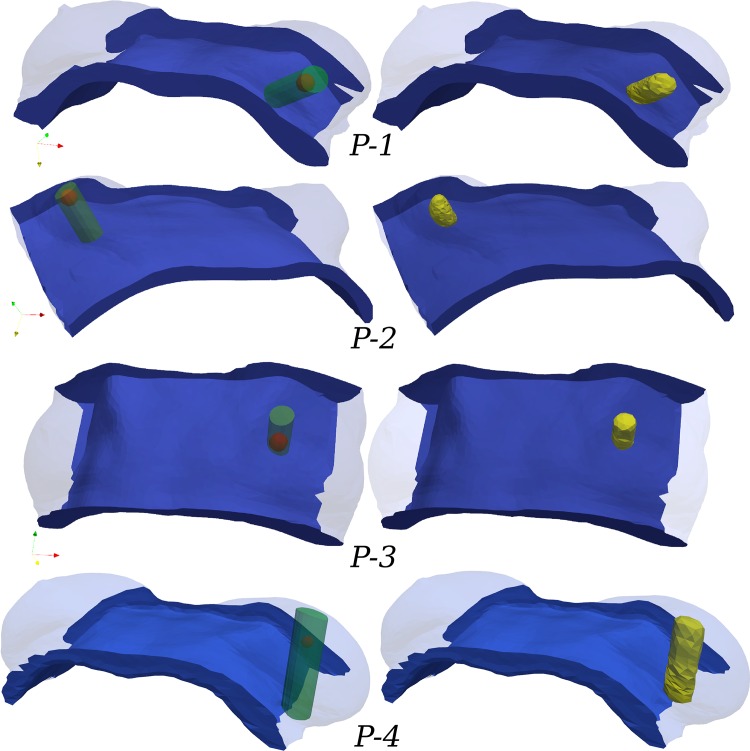
Fig 4. Adopted surgical plans in supine configuration, depicting the tumour location and the breast tissue resected.
The “idealised” surgical plan (SP) for each patient (recorded by the breast surgeon) is presented in the left column. Tumour position is shown with a red sphere and the excised breast tissues with a green transparent cylinder. In the right column, the FE models are informed by labelling the corresponding volume elements as resected tissue (shown in yellow).

