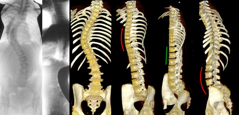
Fig 3. Posterior-anterior and lateral radiographs, and computed tomography reconstructions (OsiriX 64-bit; OsiriX Foundation, Los Angeles, CA, USA) are shown from an anterior and anterolateral view.
In this representative 15-year-old patient with adolescent idiopathic scoliosis with a typical thoracic curve pattern, anterior overgrowth cannot be observed on the lateral radiography. The rotated lordoses of both thoracic and the (thoraco)lumbar curvatures, with a clearly longer anterior spinal column than posterior elements, are shown in red, and the straight junctional segment in-between in green from a true lateral view for these regions.

