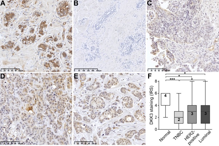
Fig 2. Loss of DKK3 protein expression in human breast cancer.
(A) Normal mammary epithelial cells showing moderate, predominantly cytoplasmic, DKK3 immunoreactivity whereas (B) primary antibody negative control is free of signal. (C-E) Weak DKK3 protein expression is observed in breast tumor samples, with lowest intensity in IHC-defined (C) TNBC cases compared to (D) HER2-positive and (E) luminal carcinomas (representative images). (F) Box plot analysis demonstrating a significant down-regulation of DKK3 in tumors of the TNBC (n = 54), the HER2-positive (n = 47) and luminal subtype (n = 362) compared to normal breast tissues (n = 11). Horizontal lines: grouped medians. Boxes: 25-75% quartiles. Vertical lines: range, minimum and maximum. * P < 0.05, *** P < 0.001, IRS: immunoreactive score.

