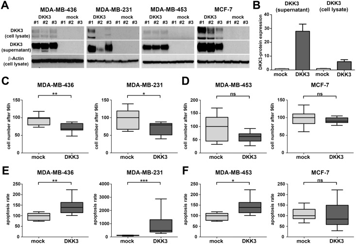
Fig 4. Stable DKK3 re-expression reduces cell growth of basal-like but not luminal-like breast cancer cell lines.
(A) Re-expression of DKK3 protein as well as secretion of DKK3 was detected by western blot. Western blot analysis was performed on total cell lysates and corresponding cell culture supernatants of three stably transfected MDA-MB-436, MDA-MB-231, MDA-MB-453 and MCF-7 DKK3 and mock clones respectively. β-actin served as a loading control. (B) All western blots depicted in A were evaluated densitometrically. In concordance, mock clones were negative for DKK3 protein whereas expression was elevated in total cell lysates of DKK3 clones. Moreover, a strong secretion of DKK3 into the cell culture supernatant could only be detected in DKK3 clones. The identical clones were used for the following functional assays. (C-D) Re-expression of DKK3 significantly reduced cell growth in basal-like (C, MDA-MB-436 and MDA-MB-231) but not luminal-like breast cancer cell lines (D, MDA-MB-453 and MCF-7). Box plots demonstrate the median cell number after 96 h cell growth of triplicate experiments. Cell growth suppression was possibly mediated by a DKK3-induced apoptosis, which was much more pronounced in breast cancer cell lines of the basal (E) than of the luminal subtype (F). Box plots demonstrate the median apoptosis rate of triplicate experiments. Horizontal lines: grouped medians. Boxes: 25-75% quartiles. Vertical lines: range, minimum and maximum. ns: not significant, * P < 0.05, ** P < 0.01, *** P < 0.001.

