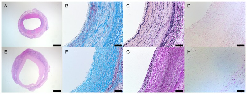
Fig 4. Histological assessment of vascular neotissue formation at 6 months.
The vascular neotissue including collagen and elastin deposition is similar to native carotid artery (CA) without ectopic calcification. Representative pictures are shown for H&E staining (A, E), Masson’s trichrome staining (B, F), Hart’s staining (C, G), and von Kossa staining (D, H). Native CA (A-D) is compared to nanofiber PCL/CS TEVGs (E-H). The scale bar represents a length of 1,000μm for A and E, and 100μm otherwise.

