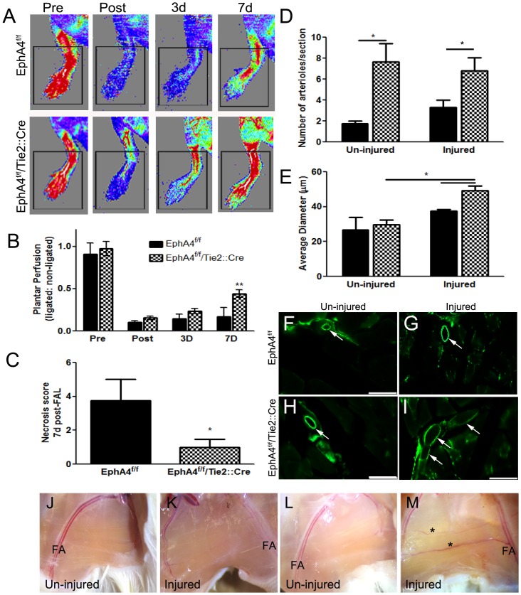
Fig 7. Plantar reperfusion and collateral remodeling post- hindlimb ischemia.
(A) Laser Doppler images of blood flow pre and post ischemia in WT and KO mice. (B) Analysis shows significant increase in plantar blood flow perfusion at 7d post hindlimb ischemia or femoral artery ligation (FAL) in KO mice compared to WT. (C) KO mice also have a significant reduction in toe necrosis compared to WT mice at 7 days post-FAL. (D) The number of CD31-positive arterioles were are in increased in serial sections of both un-injuured and injured adductor muscles in the absence of EC-specific EphA4. (E) Average arteriole diameter in the injured aductor muscle is increased in KO mice compared to WT and KO un-injured tissue at 7d post-FAL. (F-I) Representative high magnification (x20) images from un-injured and injured adductor muscles at 7d post-FAL using CD31 immuno-staining and -fluorescence. Scale bar = 100μm. (J-M) Brightfield images of WT (J and K) and KO (L and M) adductor muscles at 7d post-FAL. *P<0.05; **P<0.01; n = 5-8/group; represented as mean ± SEM. FA = Femoral artery.

