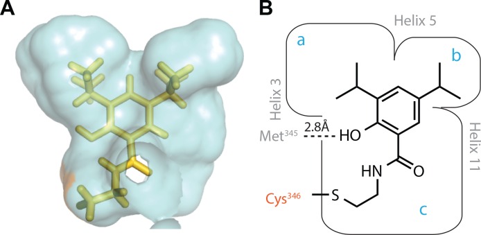
Fig 1. Computational model shows orientation of and space around top hit 15.31 in the hLRH-1 pocket.
A. Computational model of top hit 15.31 covalently bound to LRH-1 through a disulfide bond to Cys346. Van der Walls surface (blue) represents all protein within 4.5 Å of the ligand. B. Structure of 15.31 and cartoon derived from the model illustrating the binding site for 15.31 is framed by helices 3, 5 and 11 and likely includes a polar interaction with the backbone carbonyl of Met345.

