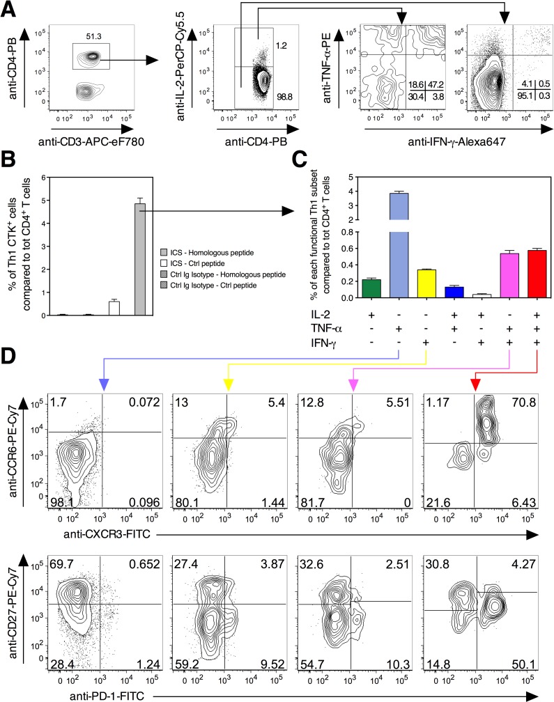
Fig 1. Cytometric strategy used to identify different functional Th1 subsets specific to mycobacterial antigens.
A) Splenocytes from C57BL/6 mice (n = 3 per group), injected s.c. with 1 x 10⁶ CFU/mouse of Mtb Δppe25-pe19, were stimulated in vitro with the PPE25:1–20 peptide at 4 weeks p.i., prior to surface and intracellular staining to detect single, double or triple positive antigen-specific Th1 cells. B) Percentage of cells producing any of the Th1 IL-2/TNF-α/IFN-γ cytokines (CTK) compared to total CD4+ T splenocytes. C) Definition of antigen-specific Th1 effector subsets as a function of their IL-2/TNF-α/IFN-γ expression and their percentages compared to total CD4+ T splenocytes. Means ± SD are standard deviations. D) Surface expression of CCR6, CXCR3, CD27 and PD-1 as analyzable in TNF-α+ or IFN-γ+ single positive, TNF-α+ IFN-γ+ double positive or IL-2+ TNF-α+ IFN-γ+ triple positive, antigen-specific functional Th1 subsets. Data are representative of two independent experiments. See furthermore S3 Fig.

