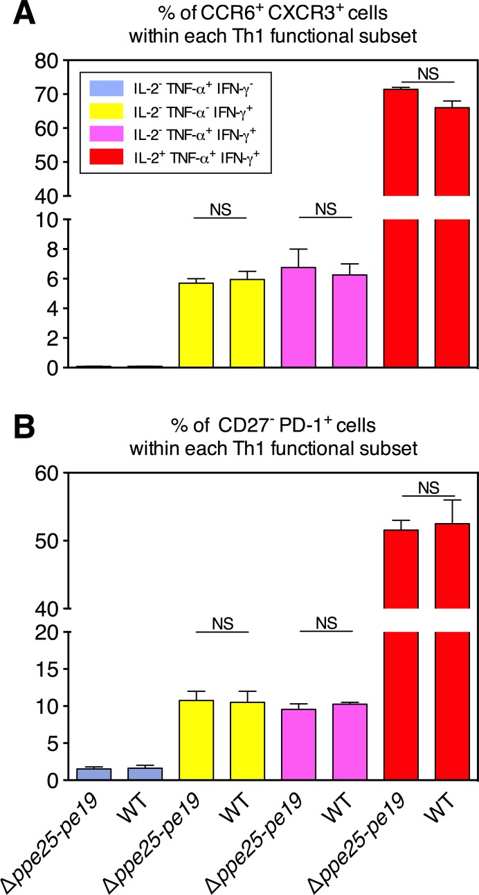
Fig 3. Comparative study of the differentiation status of the antigen-specific functional Th1 subsets in Mtb Δppe25-pe19- or Mtb WT-immunized mice.
Splenocytes from the immunized mice were stimulated with the representative PPE25:1–20 synthetic peptide as described in Materials and Methods, stained for the surface differentiation markers, and then processed for ICS specific to Th1 cytokines. Percentages of CXCR3+ CCR6+ (A) or PD-1+ CD27- (B) cells were determined, as detailed in the Fig 1D, subsequent to gating on different functional Th1 subsets. Results are means ± SD of experimental duplicates.

