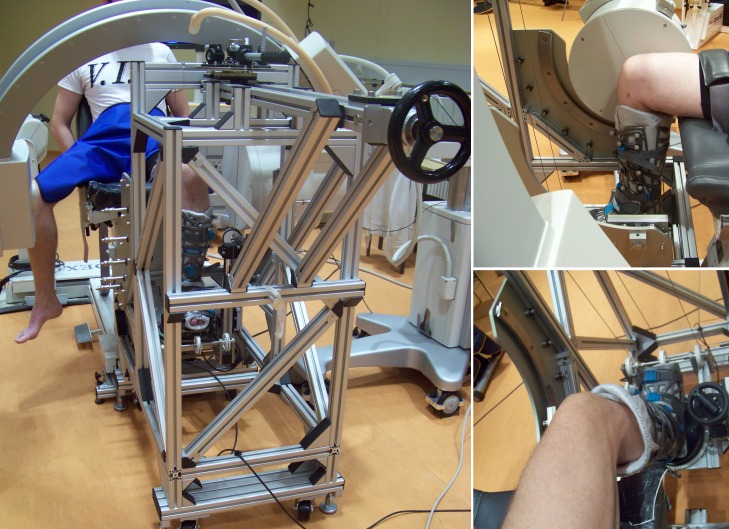
Fig 1.
Left: Measurement set-up showing a subject seated and positioned within the knee Rotometer, together with the fluoroscopic device. Top-right: Patient´s shank in the Vacoped boot and knee centred in front of the image intensifier at 90° flexion. Bottom-right: Visualization of the patient´s shank in the Vacoped boot and the attached force transducer. No artificial constraints were applied to the knee.

