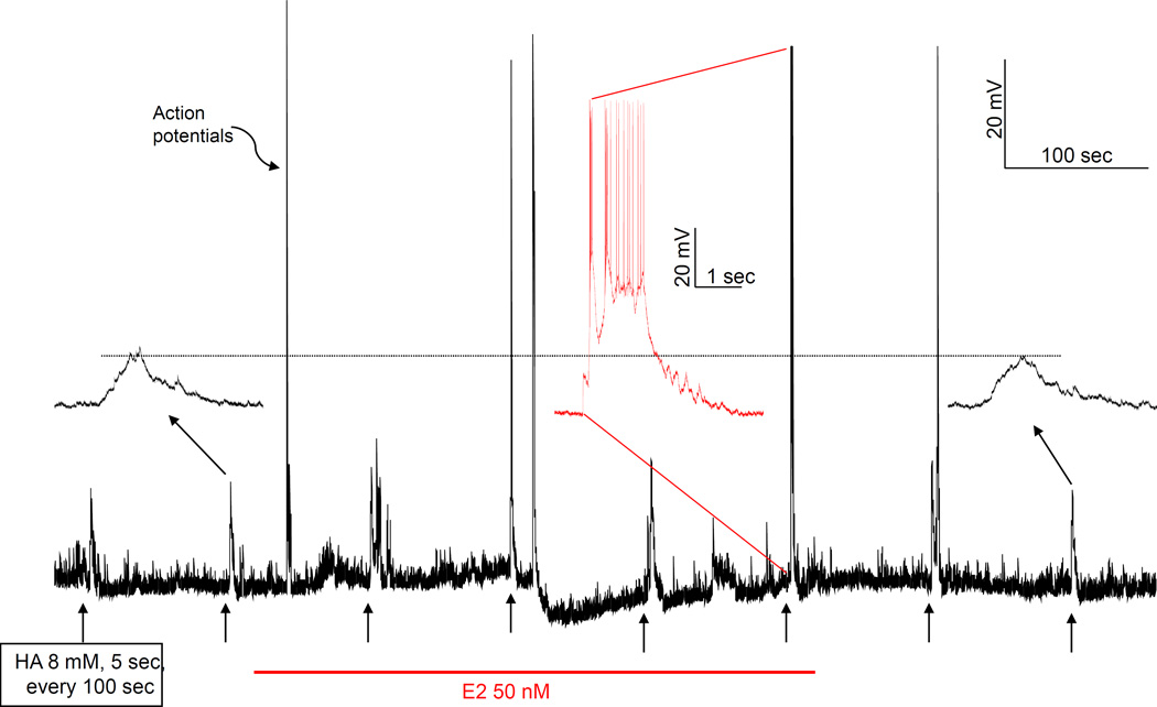
Fig. 8.
Examples of rapid E2 potentiation of depolarization induced by picospritzed HA. The trace is a continuous monitoring of the membrane potential of a VMN neuron. HA (8 mM in the ejecting pipet) was picospritzed for 5 sec every 100 sec, as indicated by short arrows below the trace. E2 was administered soon after the second HA application. In the first HA response after E2 administration, there was more activity but no action potential. By the second stimulation after E2, HA triggered action potentials and even a depolarization block that affected the next response. The second, sixth and eighth responses (indicated by longer arrows) to HA were expanded and their baseline aligned. It is clear from these insets that E2 accelerated the rate of depolarization by HA. The dashed line shows that the depolarizations induced by second and eighth HA applications were actually more depolarized than the threshold that triggered action potential in the sixth response, indicating that acute E2 lowered the threshold. Note, the neuron recovered from E2 influence gradually soon after the termination of E2 administration. Note also that E2 had no significant effect on baseline membrane potential, except for the depolarization blockade.

