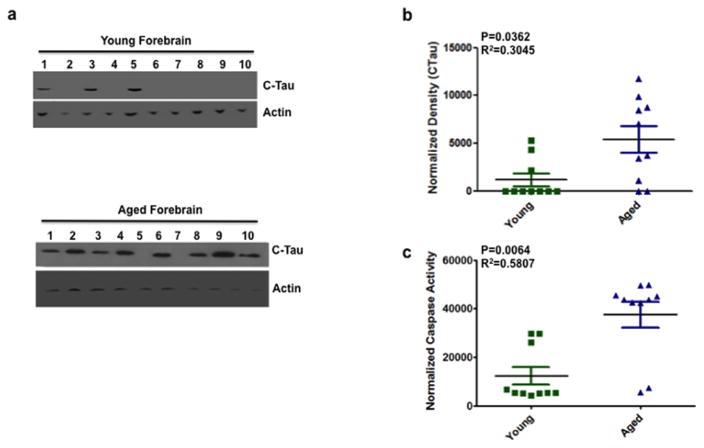
Figure 1. Increased caspase activity and cleaved tau expression in the forebrain of aged mice.
(a) Representative immunoblots from 10 animals for each group (young and aged mice forebrain) showing cleaved tau (CTau) and actin (as a loading control) expression in the forebrain. Each well number represents an individual animal. (b) Quantitative densitometry indicated that cleaved tau expression is significantly higher (P = 0.0362, R2 = 0.3045) in the forebrain of aged mice compared to young. Tau density was normalized to actin. Normalized density is given along the ordinate and the two groups (young vs. aged) are given along the abscissa. (c) DEVD-afc, a substrate used to measure caspase-3 activity indicated that caspase activity is significantly higher (P=0.0064, R2=0.5807) in the forebrain of aged mice compared to young. Caspase activity was normalized (ordinate) to buffer alone. Data are shown as mean+/−SEM. Green squares represent young individuals, blue triangles represent aged. n = 10 mice per group. Blots were confirmed in duplicate.

