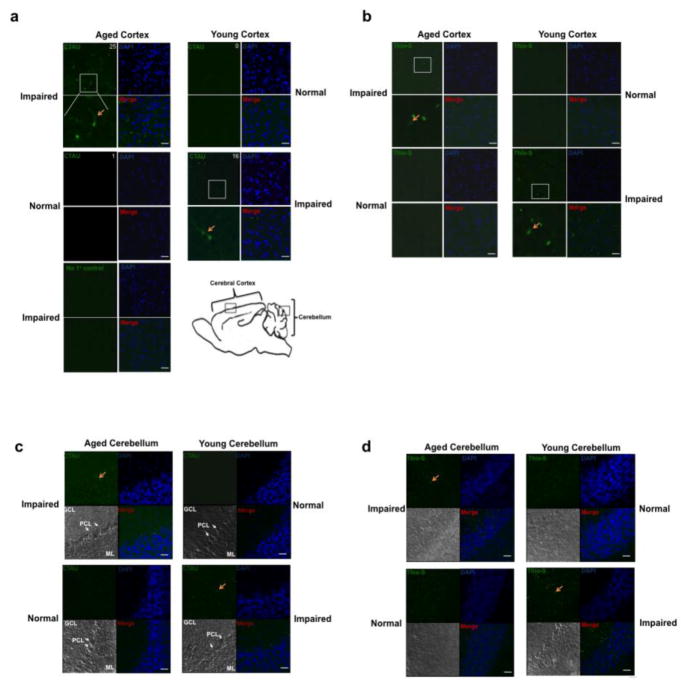
Figure 5. The aged cortex of cognitively impaired mice is positive for cleaved tau and NFTs.
(a) Aged and young forebrains were immunostained for cleaved tau (CTAU) (green). Aged mice that showed behavioral impairment also were positive for cleaved tau. Average number of cleaved TAU/area is indicated at top right of CTAU panel. (b) Similarly, the aged behaviorally impaired mice were also positive for NFTs based on Thioflavin S staining (Thio-S) (green). DAPI was used to stain nuclei (blue). Images are representative fields of the outer cortical layers of the forebrain (the region where images were taken is identified by a box in panel Af). The CTAU or Thio-S images were overlaid with the DAPI stain and shown in the merge panels, respectively. (c) Aged and young cerebellum were immunostained for cleaved tau (CTAU) (green). Aged mice showing behavioral impairment were positive for cleaved tau. (d) Similarly, aged behaviorally impaired mice were Thioflavin S positive (Thio-S) (green). Cerebellum sections were counterstained with DAPI (blue) as a nuclear marker. Red arrows indicate CTAU and Thio-S positive cells. The region where images were taken is identified by a box in panel A. Molecular layer: ML; Purkinje cell layer: PCL; Granule cell layer: GCL. Scale bars represent 25μm.

