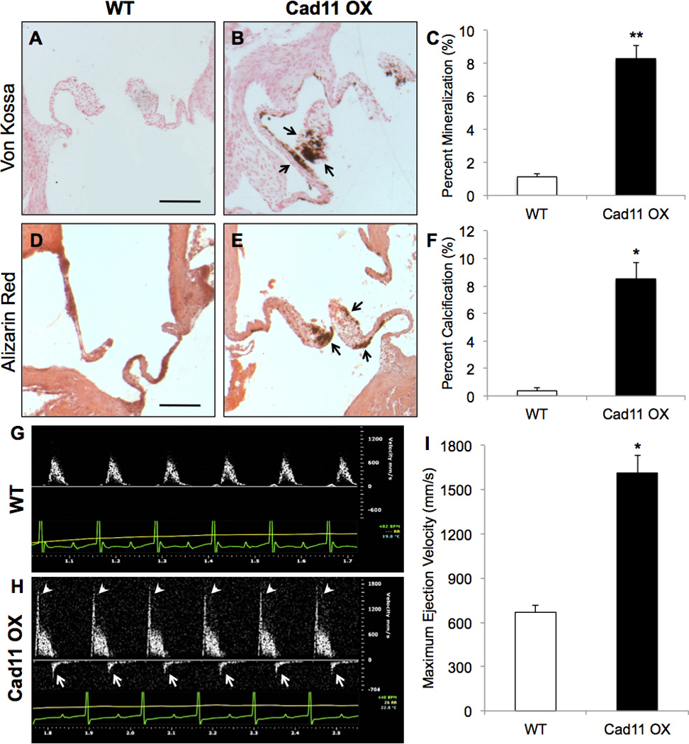
Figure 2. Cad-11 overexpression leads to formation of calcific lesions and aortic stenosis.

A–C, Von Kossa staining reveals greater mineralization (arrows, B) in adult Cad11 OX mice compared to WT mice at 10 months (n≥9, **p<8E-9) Scale bar=200μm. D–F, Alizarin Red staining reveals greater calcification (arrows, E) in adult Cad11 OX mice compared to WT mice at 10 months (n=4, *p<0.0005) G–I, Blood ejection velocity through the AoV was evaluated using Doppler ultrasound at 10 months. Arrows in H indicate regurgitation while arrowheads indicate elevated outflow velocity compared to WT mice (n=10, *p<0.0005). Significance was determined using the Student’s t-test at p<0.05.
