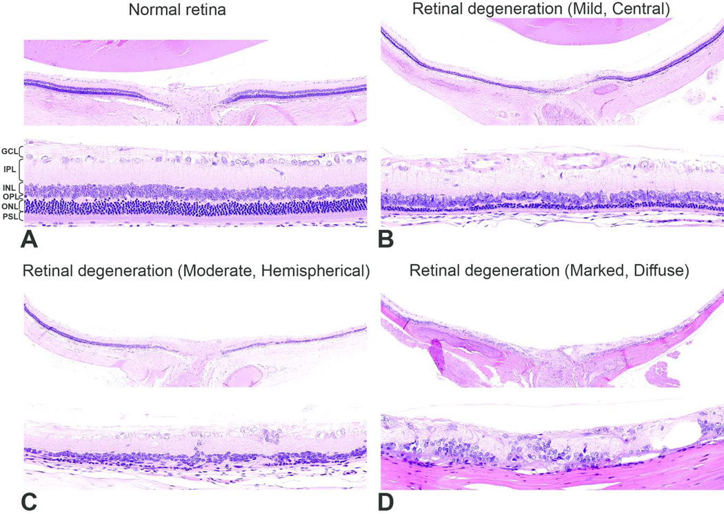
Figure 2.
Representative pictures depicting the severity of retinal degeneration. A) Grade 0 (Normal) - different retinal layers are distinct, the inner and outer segments of photoreceptor layer are elongated, outer nuclear layer is intact. B) Grade 1 (Mild) - major retinal layers remain distinct, but the photoreceptor layer and outer nuclear layer (approximately 1 to 3 rows of nuclei) are reduced in thickness. In this case, the lesion was located in the central retina. C) Grade 2 (Moderate) - photoreceptors and most of the outer nuclear layer and outer plexiform layers are lost. In this case, the lesion was located in the hemispherical retina. D) Grade 3 (Marked) - marked disruption of the normal retinal architecture with loss of normal retinal organization but with remnants of inner nuclear and ganglion cells. In this case, the lesion was diffuse. GCL, ganglion cell layer; IPL, inner plexiform layer; INL, inner nuclear layer; OPL, outer plexiform layer; ONL, outer nuclear layer; PSL, photoreceptor segment layer. H&E.

