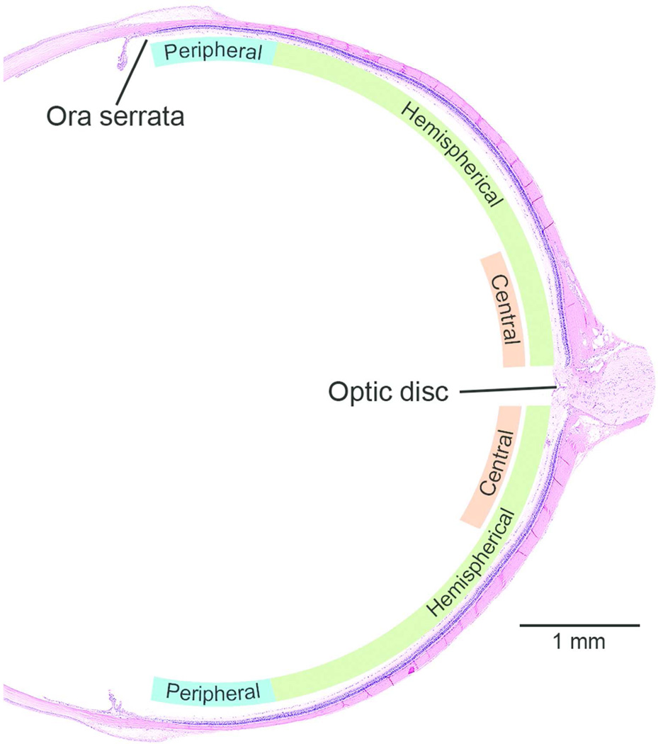
Figure 3.
Topographic localization of retinal lesions in a sagittal section. To assess whether the retinal degeneration was light-induced or not, the topography of the lesion was evaluated as follows: Diffuse - grade of the lesion is same the in the entire retina; Hemispherical - there is a distinct difference in the grade of the lesion on either side of the optic disk; Central - there is a distinct difference in the grade of the lesion just adjacent to the optic disc; and Peripheral. In general, diffuse retinal lesions are usually associated with direct chemical effect and a localized retinal lesion (at least during the early stages) may be light-induced change. Approximately 1 mm of the retina from the ora serrata was evaluated as the peripheral retina.

