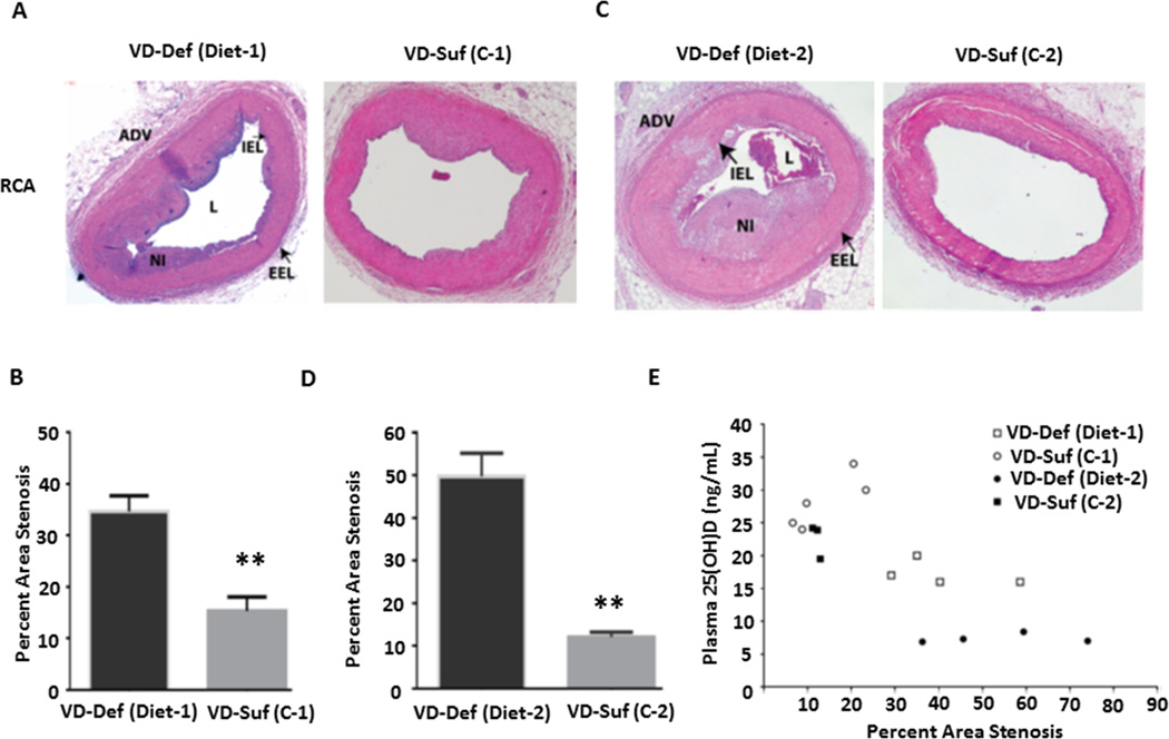
Figure 5.
Vitamin D deficiency accelerated the progression of CAD induced by hypercholesterolemic diets. The representative images of HE staining from swine right coronary arteries (RCA) are shown and the pooled data in the graph display the quantification of stenosis area in the RCAs from the swine fed on vitamin D-deficient Diet-1(VD-Def, n=4) and sufficient control (VD-Suf, C-1, n=5) diet (A and B) for one year, and vitamin D-deficient Diet-2 (n=4) and sufficient control (C-2) diet (n=3) (C and D) for one year. The relationship between plasma 25(OH)D level versus % stenosis in each swine is shown in (E). **p<0.01 vs. individual control group. ADV: adventitia, IEL: internal elastic lamina, L: lumen, EEL: external elastic lamina, NI: neointima.

