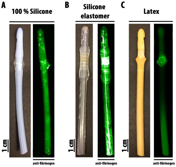
Figure 1. Representative images of the different urinary catheter materials and visualization of their fibrinogen deposition.
A) 100 % silicone; B) silicon elastomer; and C) latex. The first 10 cm of the catheter tip was used for fibrinogen deposition analysis. Deposited fibrinogen on the catheter was detected by immunofluorescence using goat anti-human fibrinogen antibody staining.

