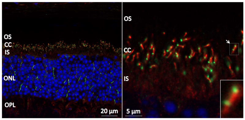
Figure 2.
CLUAP1 localizes to the connecting cilium in adult mouse retinas. Mouse immunohistochemistry staining using anti-CLUAP1 (green), anti-acetylated α-tubulin (red), and DAPI (blue). CLUAP1 can be seen to localize to specific puncti between the inner segment (IS) and outer segment (OS) layers of photoreceptors cells. CLUAP1 localization overlaps with the tip and base of the acetylated α-tubulin staining corresponding to the tip and base of connecting cilia (CC). ONL = outer nuclear layer, OPL = outer plexiform layer.

