
Figure 2.
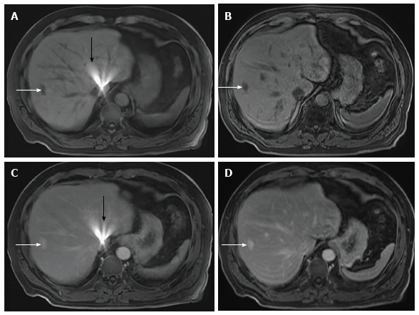
Magnetic resonance imaging of the abdomen in a 68-year-old male performed to follow-up a microcystic serous cystadenoma of the pancreas. The patient had an atrial septal defect closure device in place. Axial pre-contrast rVIBE (A), pre-contrast cVIBE (B), post-contrast rVIBE (C), and post-contrast cVIBE (D) show a hemangioma in the right liver (white arrows) with better margin delineation on pre- and post-contrast cVIBE than on pre- and post-contrast rVIBE. A radiating streak artifact in the central liver (black arrows) was seen only on pre- and post-contrast rVIBE images. VIBE: Volumetric interpolated breath-hold examination; rVIBE: Radial VIBE; cVIBE: Cartesian VIBE.