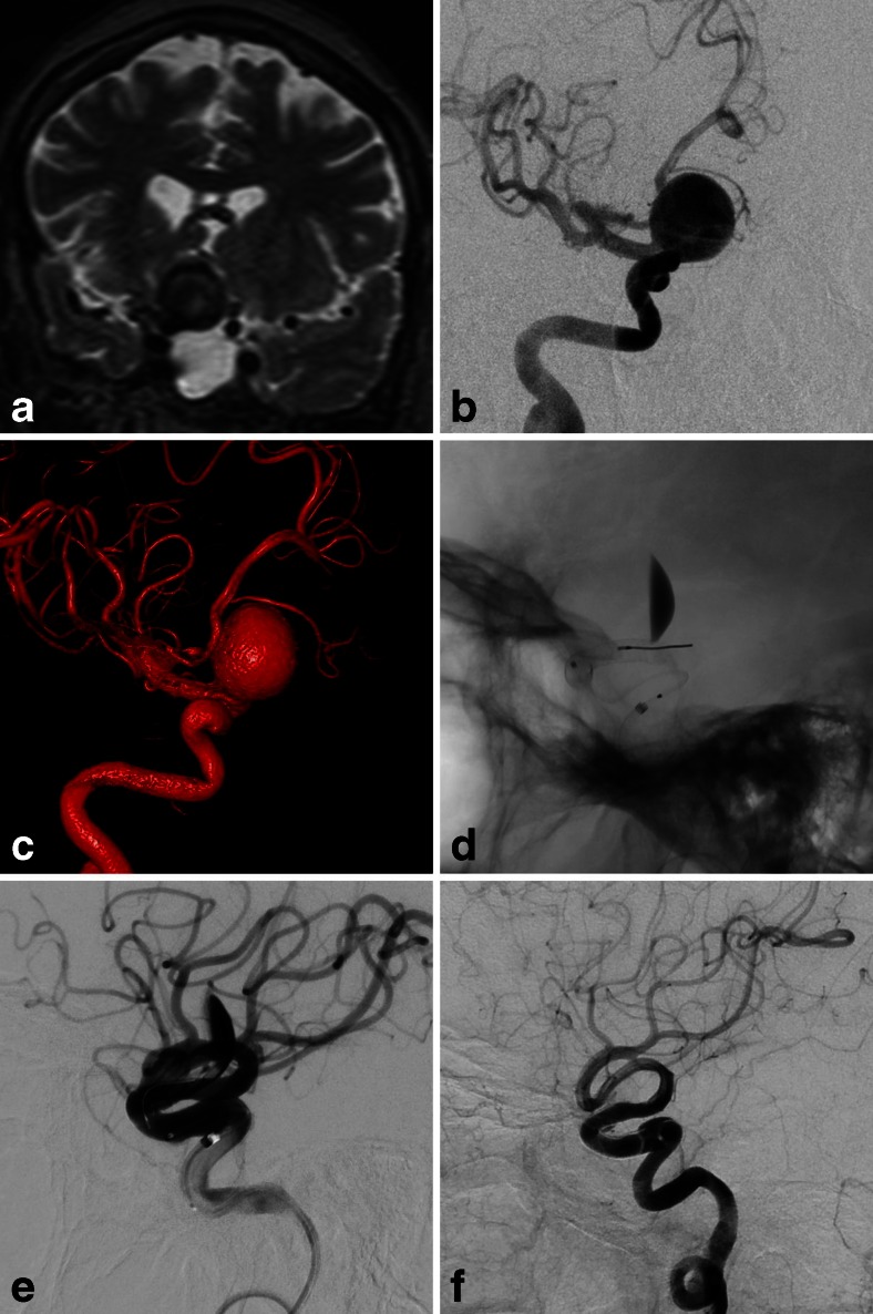
Fig. 1.
a Coronal T2-weighted magnetic resonance image of the brain shows large paraclinoid right internal carotid artery (ICA) aneurysm with mass effect on the optic chiasm. b Digital subtraction angiogram (DSA) and c 3-dimensional volume-rendered reconstruction shows wide-necked aneurysm. d Unsubtracted lateral view and e DSA of right ICA in lateral view immediately after flow diverter stent placement shows layering contrast stagnation in aneurysm and patent anterograde flow. f Six-month follow-up DSA shows vessel reconstruction with no further filling of aneurysm

