
Figure 2.
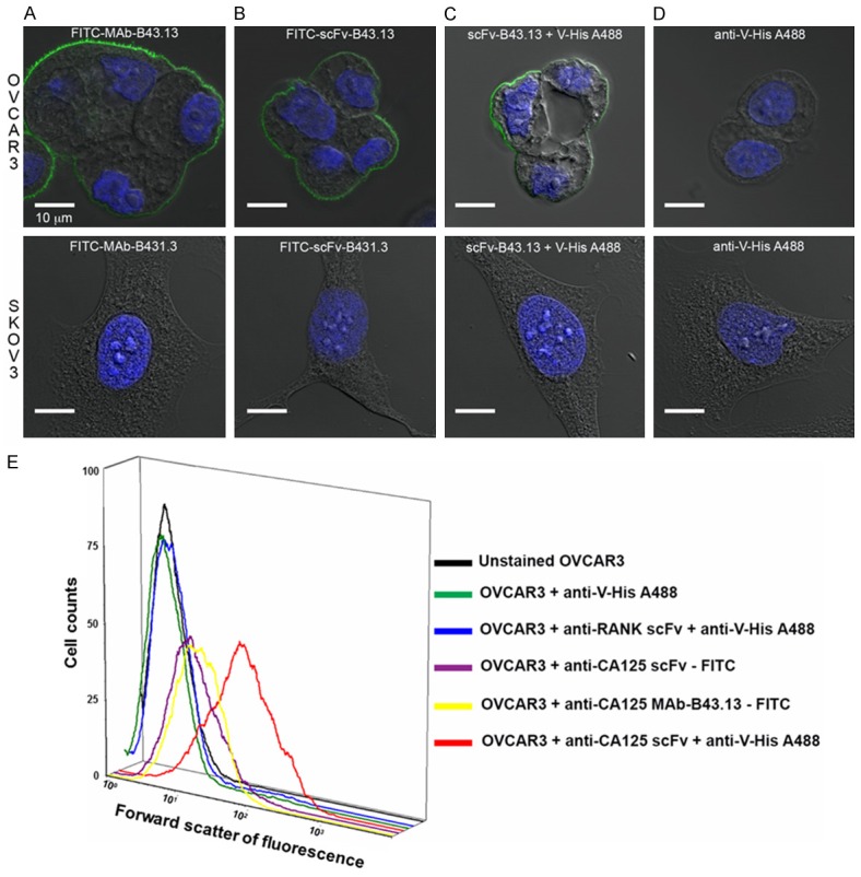
Functional Characterization of B43.13-scFv via Immunostaining and Flow Cytometry Analyses. Confocal images of OVCAR3 cells (top panel) and SKOV3 cells (bottom panel) immunostained with - A. FITC-labeled MAb-B43.13; B. FITC-labeled scFv-B43.13; C. scFv-B43.13; D. Alexa fluor 488-conjugated anti-penta histidine (V-His A488) antibody. The membrane-localized green fluorescence is a typical immunostaining pattern observed for CA125 staining in MUC16-positive cells. E. A three-dimensional histogram representation of the flow cytometry analyses of the scFv-B43.13 and control samples incubated with OVCAR3 cells. The X-axis represents forward scatter of fluorescence and Y-axis represents the cell counts.