Fig. 3.

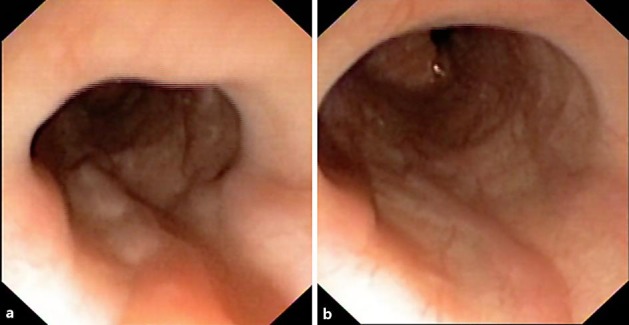
Esophagoscopy revealed two minimally protruding and slightly tortuous small varices. They showed no red wale signs (a) and were partially flattened with insufflation (b).

Esophagoscopy revealed two minimally protruding and slightly tortuous small varices. They showed no red wale signs (a) and were partially flattened with insufflation (b).