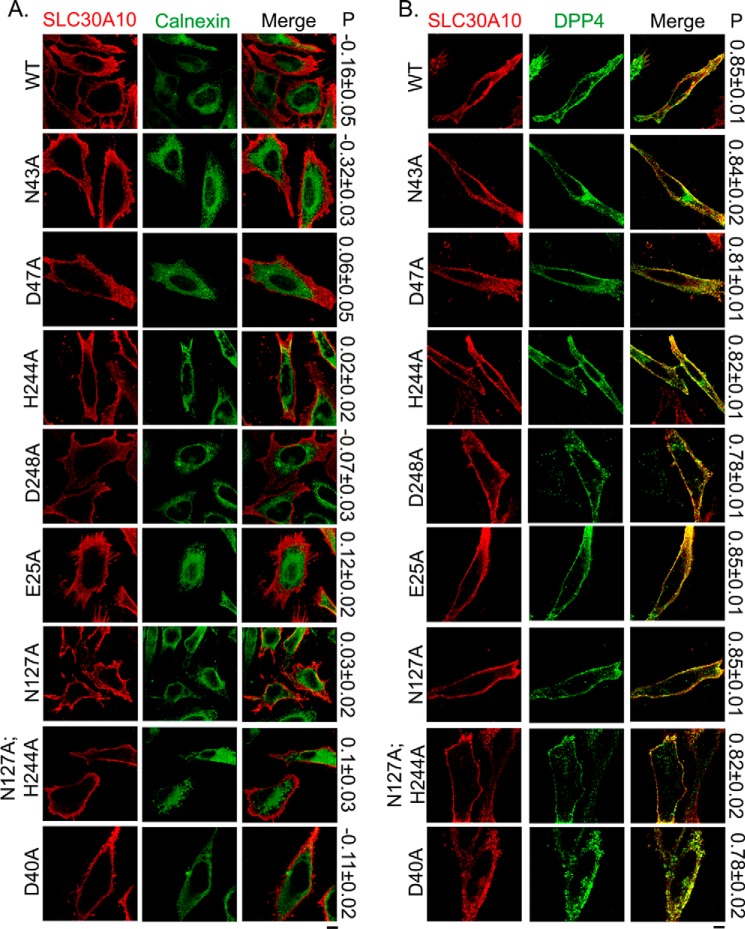
FIGURE 2.
Cell surface localization of SLC30A10WT and transmembrane domain mutants. A, HeLa cells were transfected with various FLAG-tagged SLC30A10 constructs. Two days after transfection, cultures were processed for immunofluorescence. SLC30A10 was detected using a monoclonal antibody against the FLAG epitope, and a polyclonal antibody against calnexin was used to demarcate the endoplasmic reticulum. P represents the Pearson's coefficient for colocalization between SLC30A10 and calnexin (mean ± S.E.; n = 10–15 cells per SLC30A10 construct). Scale bar, 10 μm. B, HeLa cells were cotransfected with FLAG-tagged SLC30A10WT or mutants and HA-tagged DPP4. Two days after transfection, SLC30A10 was detected using a polyclonal antibody against FLAG and DPP4 with a monoclonal antibody against HA. P denotes the Pearson's coefficient for colocalization between SLC30A10 and DPP4 (mean ± S.E.; n = 14–15 cells per SLC30A10 construct). Scale bar, 10 μm.

