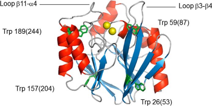
FIGURE 1.

Schematic ribbon representation of the structure of BcII 569/H/9 (Protein Data Bank code 1BVT (40)). The zinc ions at the catalytic site are represented as yellow spheres, α-helices and β-strands are shown in red and blue, respectively, and the four tryptophan residues are labeled and colored green. The β3-β4 and β11-α4 loops (residues 32–38(59–66) and 170–188(223–241), respectively) are also indicated. The figure was generated using the open-source molecular graphics system PyMOL (The PyMOL Molecular Graphics System, Version 1.2r3pre, Schrödinger, LLC).
