FIGURE 4.
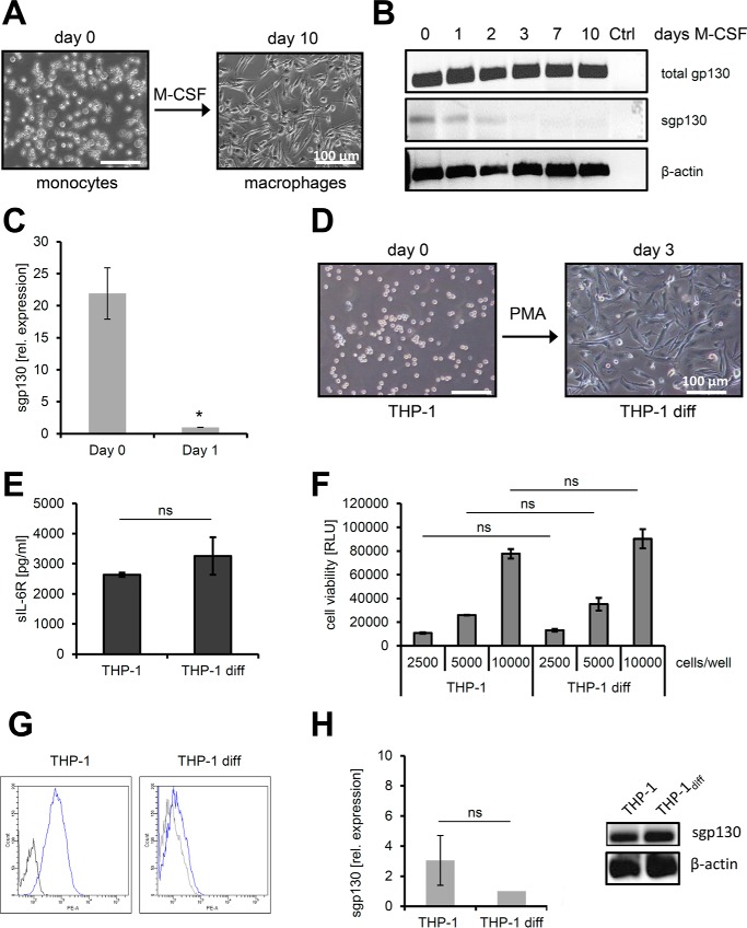
Down-regulation of full-length sgp130 during monocyte-to-macrophage transition. A, differentiation of human monocytes to macrophages was induced by addition of 100 ng/ml M-CSF for 10 days and verified by bright-field microscopy. B, semiquantitative analysis of sgp130 RNA expression in human macrophages after different time points of differentiation by RT-PCR. Total gp130 transcripts and the specific full-length variant of sgp130 were analyzed. Amplification of β-actin was used as a control (Ctrl). C, quantitative analysis of sgp130 RNA expression in human monocytes before and after 1 day of differentiation to macrophages by qPCR. Expression was normalized to GAPDH expression (mean ± S.D., n = 3). D, differentiation of the monocytic cell line THP-1 to macrophages (THP-1 diff) was induced by addition of 100 nm PMA for 3 days and verified by bright-field microscopy. E, release of sIL-6R over 72 h by monocytic THP-1 and differentiated THP-1 cells was analyzed by ELISA (mean ± S.D., n = 3). F, cell viability of 2500, 5000, or 10,000 THP-1 cells compared with the same number of differentiated THP-1 cells was analyzed as described under “Experimental Procedures” (mean ± S.D., n = 3). RLU, relative light unit. G, cell surface expression of gp130 was analyzed on THP-1 cells and on differentiated macrophages on day 3. gp130 staining is shown in blue, and control staining to exclude unspecific antibody binding is shown in black. H, analysis of sgp130 RNA expression in THP-1 cells before and differentiation (diff) by qPCR, where expression was normalized to GAPDH (mean ± S.D., n = 3), and by RT-PCR, where β-actin served as a control. If not indicated otherwise, one representative experiment of three independent experiments with similar outcomes is shown. *, p < 0.05; ns, not significant.

