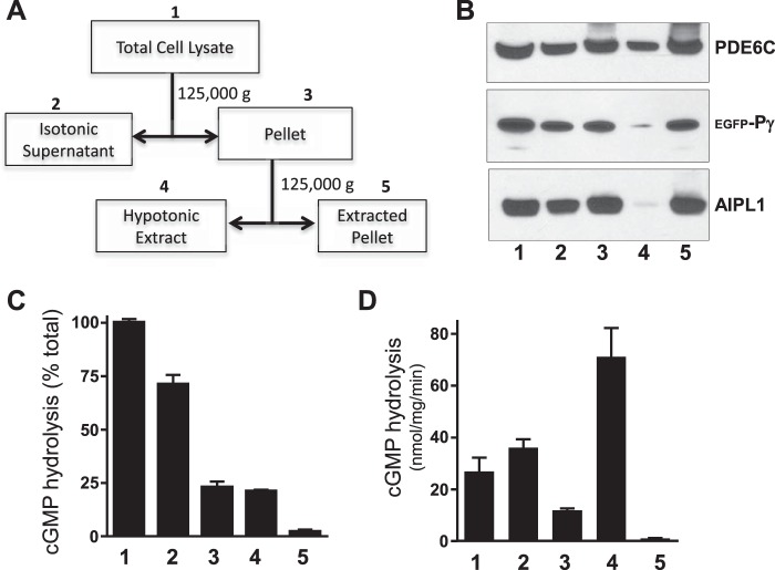
FIGURE 3.
Distribution of folded PDE6C between membrane and soluble fractions of transfected HEK293T cells. A, scheme for fractionation of lysates from HEK293T cells co-transfected with PDE6C, AIPL1 and Pγ. Details are provided under “Experimental Procedures.” B, Western blot analysis of PDE6C, AIPL1, and Pγ in the fractions obtained as outlined in A. C and D, cGMP hydrolysis in each fraction following limited trypsin treatment to remove Pγ expressed as a percentage of that in total HEK293 cell lysates (C) or activity normalized to protein content in each fraction (D) (mean ± S.E., n = 3).

