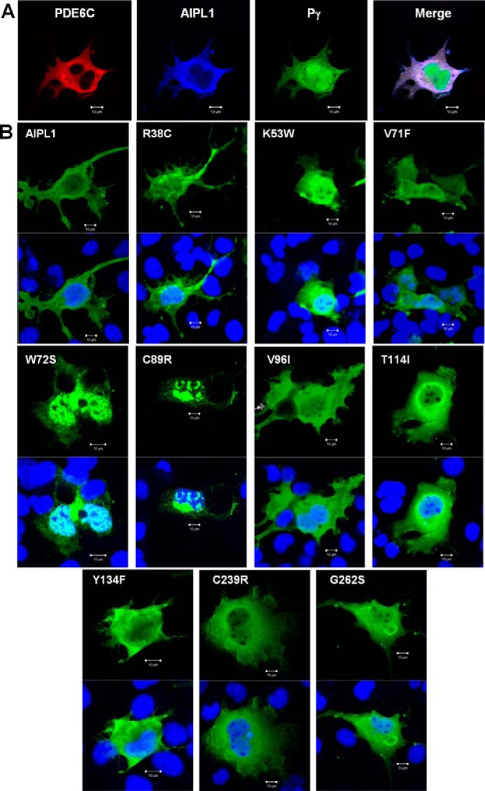
FIGURE 6.

Intracellular localization of AIPL1 mutants in COS7 cells. A, immunofluorescence images of COS7 cells co-transfected with PDE6C (red, anti-PDE6C), AIPL1 (blue, anti-HA), and Pγ (green, EGFP fluorescence). B, immunofluorescence images of COS7 cells transfected with AIPL1 mutants (AIPL1, green; TO-PRO3 nuclear stain, blue). Cells transfected with the AIPL1 mutant proteins W72S and C89R show formation of nuclear and perinuclear inclusion bodies.
