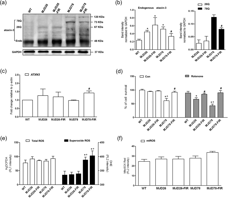
Figure 1. Expression of ataxin-3 with non-pathologic and pathologic polyQ, cell viability and oxidative stress in MJD cells after 3-day FIR treatment.
(a) Western blot analyzed the protein expressions of ataxin-3 including (b) the endogenous ataxin-3 and the polyQ expansion of 26 (26Q) or 78 CAG-repeat expansion (78Q) were quantified in human neuroblastoma cells (SK-N-SH) overexpressing 26- (26Q, internal control, MJD26) and 78-CAG repeats (MJD78) in ATXN3 gene with or with FIR treatment. The protein quantification was calculated by three-time independent analysis at least. (c) The mRNA level of ATXN3 was detected by using the RT-PCR with the primers flanking non-GAC repeat regions. (d) Cell viability and sensitivity of rotenone (100 nM)-induced cell death was evaluated by PI staining using a flow cytometry. The oxidative stress was comprehensively assessed by flow cytometry analysis of (e) intracellular (Total) ROS (H2DCFDA staining), superoxide (DHE staining) and (f) mitochondrial superoxide (mtROS, MitoSOX Red staining). *p < 0.05, compare to WT group; #p < 0.05, compare to each non-treated group of MJD cells; +p < 0.05, compare to non-treated MJD26 group.

