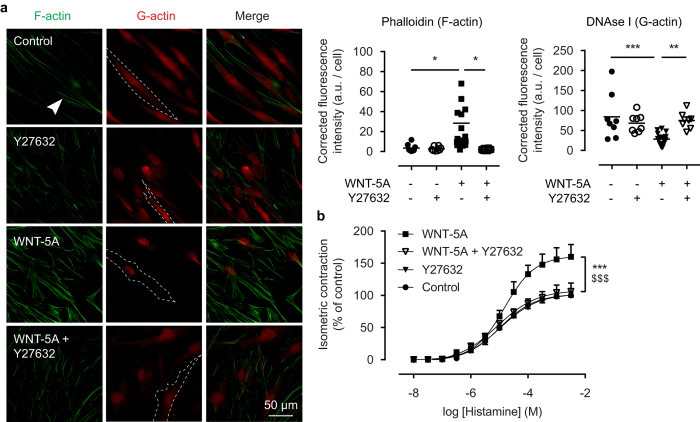
Figure 4. ROCK activation underlies WNT-5A-induced actin polymerisation.
(a) Representative immunofluorescent images of a Phalloidin (F-actin, green) and DNAse I (G-actin, red) staining of airway smooth muscle cells exposed to WNT-5A (200 ng/mL) for 2 hours in the presence or absence of Y27632 (1.0 μM), and the corresponding quantification. White arrowhead points to filamentous actin. Dashed line represents a single cell boundary. Horizontal line represents the mean. (b) Organ-cultured bovine tracheal smooth muscle strips were pre-incubated with WNT-5A (500 ng/mL) and/or Y27632 (1.0 μM) for 48 hours and a cumulative dose-response curve to histamine for the maximum isometric tension was constructed. *vs Control, $ vs WNT-5A + Y27632. Data represents five independent experiments, each performed in duplicate. Data is expressed as the mean ± SEM.

