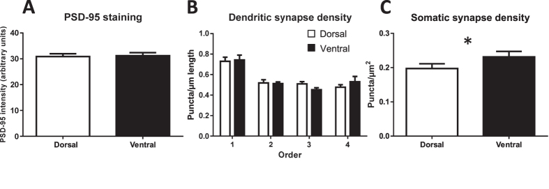
Figure 7. Septotemporal characterization of synapse-related immunoreactivity.
(A) Synapse-related immunoreactivity in the molecular layer surrounding the DG does not differ between DG subregions along the septotemporal axis. (B) Dendritic synapse-related puncta density does not differ for immature neurons in the dorsal versus ventral DG; overall, density is highest in the first dendritic branch. (C) Somatic synapse-related puncta density is higher for immature neurons in the ventral DG. *p ≤ 0.05.

