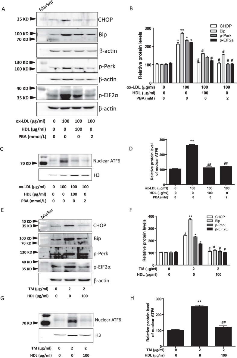
Figure 5. HDL suppresses ER stress response in 3T3-L1 adipocytes induced by ox-LDL or TM.
(A–D) Adipocytes were pretreated with HDL or PBA in DMEM with 1% FBS for 2 hr, followed by incubation with ox-LDL and Sandoz58035 (10 μg/ml) for 48 hr, and then (A,B) the protein levels of ER stress markers were evaluated by Western blot. (C,D) The levels of ATF6 protein in nuclear were analyzed by Western blot and normalized to histone H3 level. (E–H) Adipocytes were pretreated with or without HDL for 2 hr, and then followed by incubation with TM and sandoz58035 for 24 hr, and then protein levels of ER stress markers and nuclear ATF6 were determined by Western blot. Data are expressed as the mean ± SEM of at least four independent experiments. *P < 0.05, **P < 0.01 versus control group; #P < 0.05, ##P < 0.01 versus ox-LDL or TM group.

