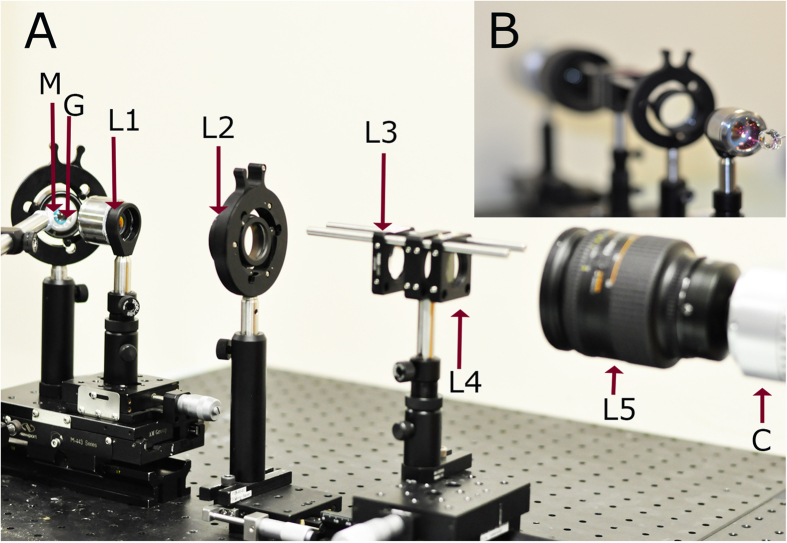
Figure 1. Optical setup of developed indirect gonioscopic imaging system.
(a) Optical setup of the developed imaging system. (b) Inset, illustrating the front view of our system. [M: Eye model; G: Hoskins-Barkan Goniotomy lens; L1: objective lens; L2: tube lens; L3: convex lens; L4: axicon lens; L5: zoom lens; C: CCD].

