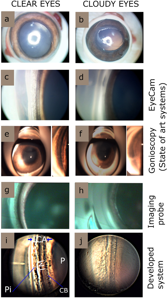
Figure 7. Comparison of various imaging system for imaging iridocorneal angle for clear and cloudy eyes.
(a,b) Example photographs of clear and cloudy eyes respectively. (c,d) Still images by EyeCam (Clarity Medical systems, Pleasanton, CA, USA). (e,f) Still images through Gonioscope (Latina 5 Bar SLT, single mirror, Ocular instruments, WA, USA). (g,h) Still images aptured using dual modality hand held probe. (i,j) Still images taken by our imaging system. Note for (e–h) the angle regions of gonioscopy images are digitally zoomed and highlighted as separate insets. [ICA: iridocorneal angle; P: Pupil; I: Iris; Pi: Pigmentation CB: Ciliary body, TM: Trabecular meshwork.]

